At a particular connection in the hippocampus, a nerve cell releases a chemical that helps restrain its neighbor. A few moments later, an impulse reaches the same nerve ending again. The cells are still connected. Yet the second impulse may release less of the restraining chemical than the first. To understand why, it helps to look at what the receiving cell did in the interval.
It was once tempting to draw a synapse with a single arrow. One side sends a chemical messenger, which crosses a narrow gap and acts on the other side. The arrow is accurate as far as it goes. Some receiving neurons can also release a short-lived chemical of their own that travels back to the sending terminal. Among the best-studied messengers for this return trip is an endocannabinoid called 2-AG.
Start with the forward signal
Consider a connection between an inhibitory nerve cell and a pyramidal neuron in the hippocampus. When an electrical impulse arrives at the inhibitory cell’s terminal, it can trigger the release of GABA. This neurotransmitter crosses the gap and acts on receptors on the pyramidal neuron. Under the usual adult conditions, the effect helps restrain that neuron’s activity. Each impulse has a certain probability of releasing transmitter.
Now let the receiving neuron become strongly active. A rise in calcium inside it can help set in motion the production of an endocannabinoid from lipids in the cell membrane. At many synapses the crucial molecule is 2-arachidonoylglycerol, hence 2-AG. As Masanobu Kano explains in a review of the underlying physiology, particular receptors on the receiving neuron can also help trigger the process. The chemical message reflects something that has just happened on the receiving side.
The return address is on the terminal
The released 2-AG can reach a receptor called CB1 on the sending terminal. Activation of that receptor reduces the probability of transmitter release. The next incoming impulse may consequently deliver a smaller GABA signal to the pyramidal neuron. This temporary reduction in inhibition is sometimes called disinhibition. It can make the receiving neuron easier to excite even though the direct action at the terminal was to suppress chemical release.
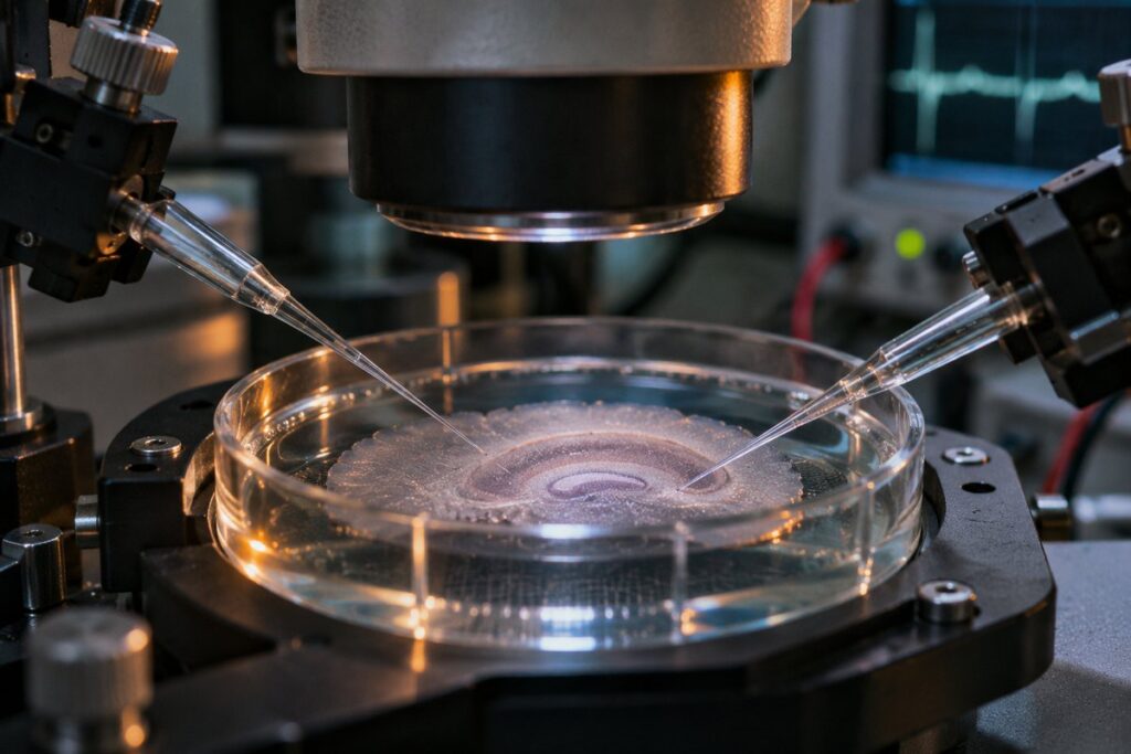
In a classic hippocampal experiment, Rachel Wilson and Roger Nicoll investigated the brief reduction in inhibitory transmission that follows electrical depolarization of a receiving neuron. Their results implicated cannabinoid receptors in the return signal. That electrical record offered something that a chemical diagram alone could never supply. It showed when changing the receiving cell altered what came back to it from its neighbor.
How do you identify a messenger you barely see?
Discovering which substance carried the message took more than locating the receptor. Several molecules made by the body can activate cannabinoid receptors. Anandamide, which researchers had isolated from brain tissue years earlier, was an obvious candidate. Pulling a molecule out of an extract, however, doesn’t show that a particular pair of cells uses it at a particular instant.
One line of evidence came from taking away an enzyme. In experiments with genetically modified mice, researchers examined what happened when neurons lacked a major enzyme for making 2-AG. The familiar retrograde suppression disappeared in the synapses they studied. The result tied the electrical behavior much more tightly to the production of a specific lipid messenger.
It is worth returning to the two impulses that opened the story. If the original terminal had been releasing an excitatory transmitter, reducing its release would have a different consequence for its neighbor. And a terminal without the relevant receptor would not receive this particular chemical reply. A brain-wide description such as “cannabinoids slow things down” loses both those details. At an individual synapse the question has a sharper answer. Which cell spoke first, what did it release, and which of the two cells had something to say back?